Critical information on fetal well-being collected longitudinally across pregnancy.
Serial images are taken by a dedicated study radiologist on a GE Voluson E8 Expert (General Electric Healthcare, Chicago, Illinois) ultrasound machine at the study site.
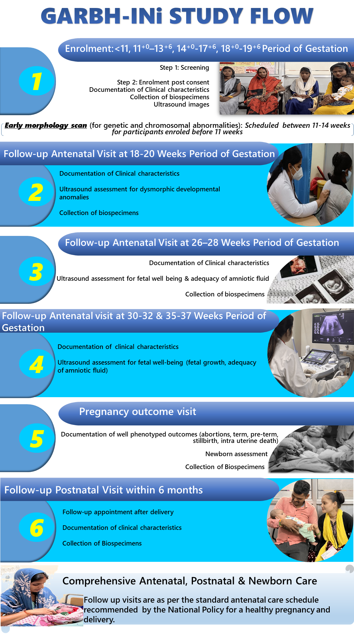
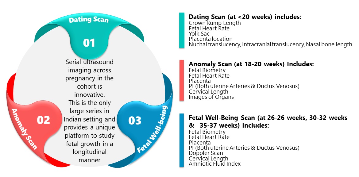
Abdominal ultrasound scans are performed to assess Period of Gestation (POG), fetal growth & well-being, placental location & morphology, as well as the morphological markers for genetic and chromosomal abnormalities at 11–14 weeks. Dysmorphic developmental anomalies are evaluated by ultrasound scans done at 18–20 weeks.
Serial changes in cervical length are monitored using transvaginal scans.
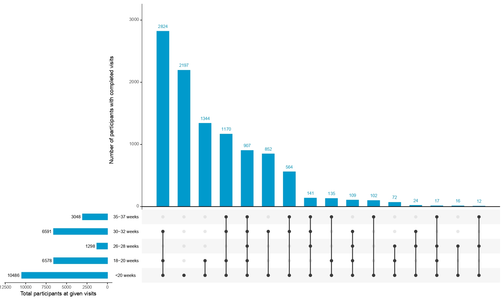
Doppler scans scheduled between 26-28 weeks, 30 and 32 weeks and 35-37 weeks POG evaluates uterine and umbilical artery pulsatility indices and amniotic fluid index as markers of fetal wellbeing; fetal middle cerebral artery and ductus venosus blood flow are measured as potential markers of fetal growth restriction.
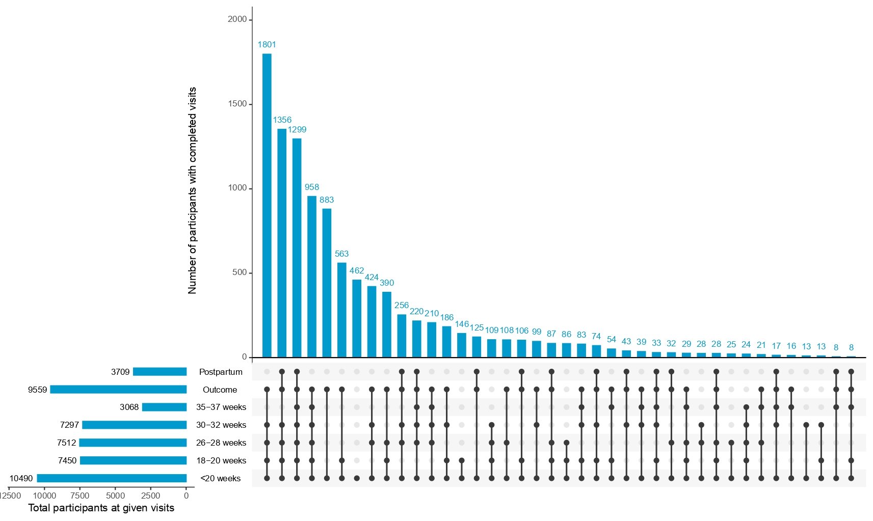
Images available
<11 weeks, 11-14 weeks, 14-18 weeks, 18-20 weeks, 26-28 weeks, 30-32 weeks, 35-37 weeks
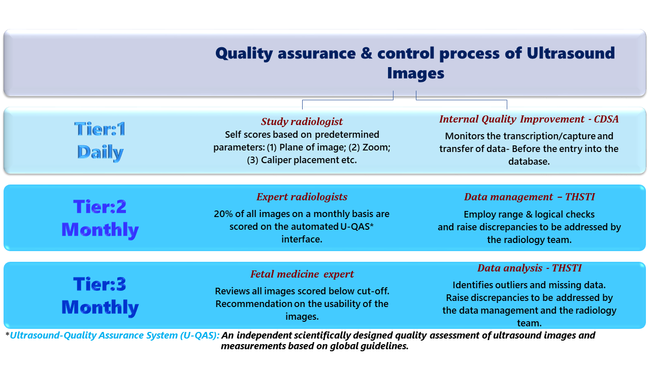
Quality control is ensured through standardized protocols, monthly review of images by experienced radiologists and training and retraining of the study radiologists.
Coordination for submission of de-identified ultrasound imaging data with IBDC ongoing
Explore Ultrasound Variables